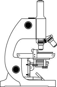
42 compound microscope diagram without labels
Labeling the Parts of the Microscope | Microscope World Resources Labeling the Parts of the Microscope This activity has been designed for use in homes and schools. Each microscope layout (both blank and the version with answers) are available as PDF downloads. You can view a more in-depth review of each part of the microscope here. Download the Label the Parts of the Microscope PDF printable version here. Label A Microscope Teaching Resources | Teachers Pay Teachers This is a set of 3 tiered readings. Students will read a passage about the how to use a compound light microscope. Students will use textual evidence to answer questions and label the different parts of the microscope. It also allows students to gain prior knowledge about the compound microscope. Version A provides the most support for students.
› 48903430 › Inorganic_Chemistry_4Inorganic Chemistry 4th edition, Catherine Housecroft Enter the email address you signed up with and we'll email you a reset link.

Compound microscope diagram without labels
Compound Light Microscope Diagram Worksheet Modern compound light microscopes under optimal conditions can we an average from 1000X to 2000X times the specimens original diameter Diagram. Label the parts of the microscope using the word... Label the microscope — Science Learning Hub In this interactive, you can label the different parts of a microscope. Use this with the Microscope parts activity to help students identify and label the main parts of a microscope and then describe their functions. Drag and drop the text labels onto the microscope diagram. Labelled Diagram of Compound Microscope - Biology Discussion The below mentioned article provides a labelled diagram of compound microscope. Part # 1. The Stand: The stand is made up of a heavy foot which carries a curved inclinable limb or arm bearing the body tube. The foot is generally horse shoe-shaped structure (Fig. 2) which rests on table top or any other surface on which the microscope in kept.
Compound microscope diagram without labels. Parts of a microscope with functions and labeled diagram - Microbe Notes Figure: Diagram of parts of a microscope There are three structural parts of the microscope i.e. head, base, and arm. Head - This is also known as the body. It carries the optical parts in the upper part of the microscope. Base - It acts as microscopes support. It also carries microscopic illuminators. A Study of the Microscope and its Functions With a Labeled Diagram ... Compound Microscope Diagram The compound microscope uses light for illumination. Some compound microscopes make use of natural light, whereas others have an illuminator attached to the base. The specimen is placed on the stage and observed through different lenses of the microscope, which have varying magnification powers. Parts of a Compound Microscope and Their Functions - NotesHippo Compound microscope magnification is determined by multiplying the eyepiece and objective powers. When viewed through a 5X eyepiece with a 10X objective, an item is magnified 5 x 10=50 times. The magnification is 10 x 45 = 450 times when using a 10X eyepiece and a 45X objective. How to Use the Compound Microscope Compound Microscope- Definition, Labeled Diagram, Principle, Parts, Uses Alternatively, the magnification of the compound microscope is given by: m = D/ fo * L/fe where, D = Least distance of distinct vision (25 cm) L = Length of the microscope tube fo = Focal length of the objective lens fe = Focal length of the eye-piece lens Parts of a Compound Microscope Eyepiece And Body Tube.
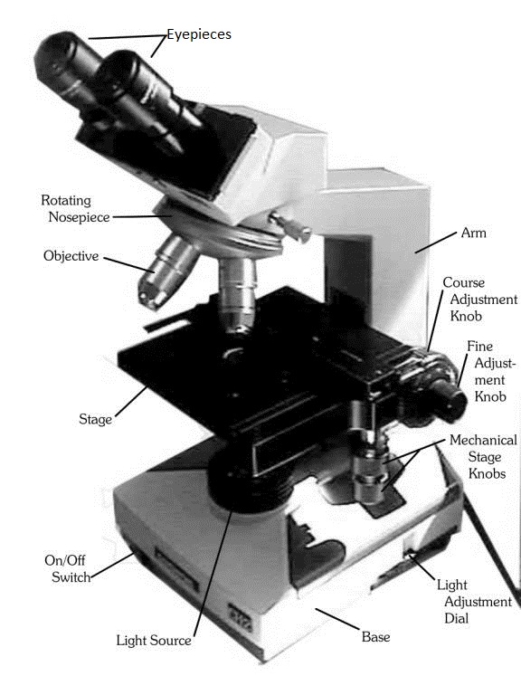
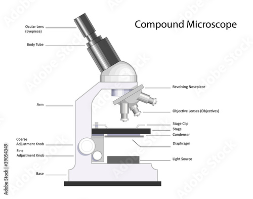
Working Principle and Parts of a Compound Microscope (with Diagrams) It holds the stage, body tube, fine adjustment and coarse adjustment. 5. Body Tube: It is usually a vertical tube holding the eyepiece at the top and the revolving nosepiece with the objectives at the bottom. The length of the draw tube is called 'mechanical tube length' and is usually 140-180 mm (mostly 160 mm). 6. Compound Microscope: Definition, Diagram, Parts, Uses, Working ... - BYJUS A compound microscope is defined as A microscope with a high resolution and uses two sets of lenses providing a 2-dimensional image of the sample. The term compound refers to the usage of more than one lens in the microscope. Also, the compound microscope is one of the types of optical microscopes. Compound Microscope Parts, Functions, and Labeled Diagram Compound Microscope Definitions for Labels. Eyepiece (ocular lens) with or without Pointer: The part that is looked through at the top of the compound microscope. Eyepieces typically have a magnification between 5x & 30x. Monocular or Binocular Head: Structural support that holds & connects the eyepieces to the objective lenses. Compound Microscope: Parts of Compound Microscope - BYJUS (A) Mechanical Parts of a Compound Microscope 1. Foot or base It is a U-shaped structure and supports the entire weight of the compound microscope. 2. Pillar It is a vertical projection. This stands by resting on the base and supports the stage. 3. Arm The entire microscope is handled by a strong and curved structure known as the arm. 4. Stage
16 Parts of a Compound Microscope: Diagrams and Video Once you have an understanding of the parts of the microscope it will be much easier to navigate around and begin observing your specimen, which is the fun part! The 16 core parts of a compound microscope are: Head (Body) Arm Base Eyepiece Eyepiece tube Objective lenses Revolving Nosepiece (Turret) Rack stop Coarse adjustment knobs Compound Microscope - Types, Parts, Diagram, Functions and Uses A compound microscope captures an inverted image of the specimen because every time the light passes through the lens, the image's direction is flipped. The image always ends up inverted from the original. So, if you move the sample to the left, it moves in the right direction. Image 18: A comparison image between a simple and compound microscope. Parts of the Microscope with Labeling (also Free Printouts) 5. Knobs (fine and coarse) By adjusting the knob, you can adjust the focus of the microscope. The majority of the microscope models today have the knobs mounted on the same part of the device. Image 5: The circled parts of the microscope are the fine and coarse adjustment knobs. Picture Source: bp.blogspot.com. The Microscope- compound microscope diagram - Major Science and technology The invention of the microscope is unknown. It is believed that Zacharias Janssen and his father Hans were responsible for making the first mixed microscope in the Netherlands in the latter part of the 16th century.Galileo is sometimes listed as the inventor, but this is unlikely to be true. In 1665 Robert Hooke produced a very influential book called Microstria in which he drew images of ...
Microscope With Labels Clip Art at Clker.com - vector clip art online, royalty free & public domain
PDF Label compound microscope worksheet - Weebly [clearBoth] [clearBoth] Microscope diagram without label After you've studied all the pieces of the composite microscope, it's time to put your brain to the test. Print an unmarked microscope chart and check that you can fill out all the blanks. [clearBoth] [clearBoth] Blank microscope diagram Next we have an empty microscope diagram.

Compound Microscope Labeling Worksheet - worksSheet list
› 37409050 › general_chemistry_pdf(PDF) general-chemistry.pdf | Sumit Banerjee - Academia.edu Enter the email address you signed up with and we'll email you a reset link.

Microscope Labelled Gcse - Micropedia
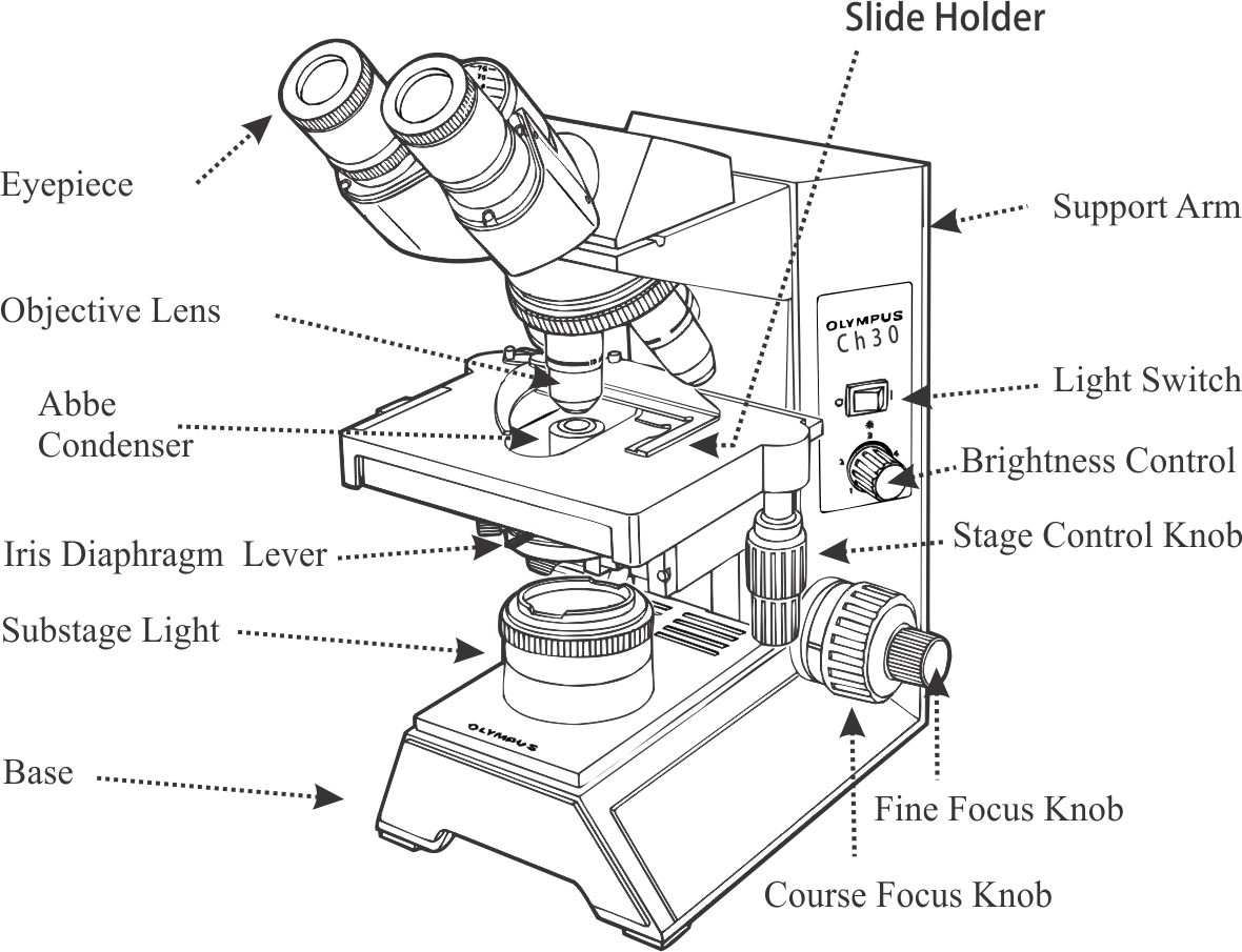
PDF The Compound Light Microscope - teachers.wrdsb.ca The Compound Light Microscope TASK Refer to page 605 in your text to: 1. Name each of the structures described in the table to the right. 2. Match each structure to the letter in the diagram below. ** ALWAYS USE TWO HANDS TO CARRY A MICROSCOPE** Letter Structure Function joins body tube to base supports the entire microscope
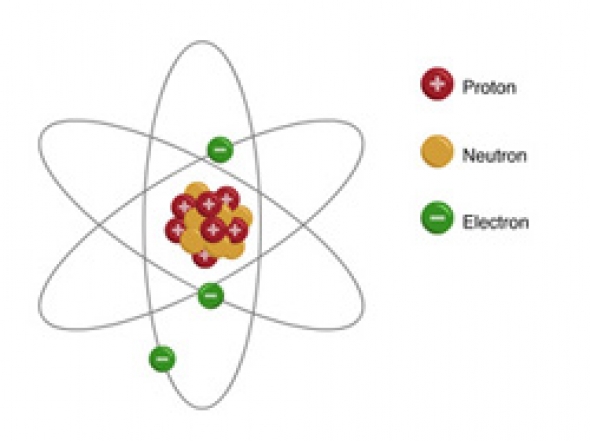
can i get a structure of an atom with labellings Science Materials Metals and Non-Metals ...
Label Microscope Diagram - EnchantedLearning.com Using the terms listed below, label the microscope diagram. arm - this attaches the eyepiece and body tube to the base. base - this supports the microscope. body tube - the tube that supports the eyepiece. coarse focus adjustment - a knob that makes large adjustments to the focus. diaphragm - an adjustable opening under the stage, allowing ...

Compound light microscope diagram. | Mad scientist | Pinterest | Cell structure, Biology and Science
en.wikipedia.org › wiki › FluorescenceFluorescence - Wikipedia The chemical compound responsible for this fluorescence is matlaline, which is the oxidation product of one of the flavonoids found in this wood. [1] In 1819, Edward D. Clarke [5] and in 1822 René Just Haüy [6] described fluorescence in fluorites , Sir David Brewster described the phenomenon for chlorophyll in 1833 [7] and Sir John Herschel ...
10 Best Images of Microscope Drawing Worksheet - Microscope Parts Labeled, Plant and Animal Cell ...
› biopython › biopython_quickBiopython - Quick Guide - tutorialspoint.com Create a Track for each track you want on the diagram, and add GraphSets and FeatureSets to the tracks you require. Create a Diagram, and add the Tracks to it. Tell the Diagram to draw the image. Write the image to a file. Let us take an example of input GenBank file −

Heys~
en.wikipedia.org › wiki › Electron_microscopeElectron microscope - Wikipedia An electron microscope is a microscope that uses a beam of accelerated electrons as a source of illumination. As the wavelength of an electron can be up to 100,000 times shorter than that of visible light photons, electron microscopes have a higher resolving power than light microscopes and can reveal the structure of smaller objects.
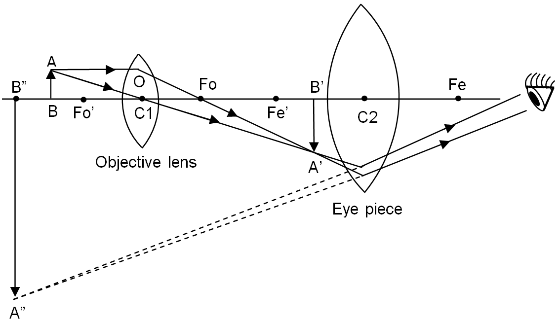
Draw a labelled ray diagram of a compound microscope and explain its working
PDF Parts of a Microscope Printables - Homeschool Creations typical student microscope -other microscopes will vary) •Which part of the microscope rotates so another person can look through the eyepiece without needing to move the microscope ? the head •What is the magnification level on the eyepiece of a microscope?10x (see objective lens magnification to see how these work together)

Compound Microscope Diagram Blank - Diagram Media
Parts of a Compound Microscope (And their Functions) - Scope Detective List of Microscope Parts and their Functions. 1. Ocular Tubes (Monocular, Binocular & Trinocular) The ocular tubes, are to tubes that lead from the head of the microscope out to your eyes. On the end of the ocular tubes are usually interchangeable eyepieces (commonly 10X and 20X) that increase magnification.
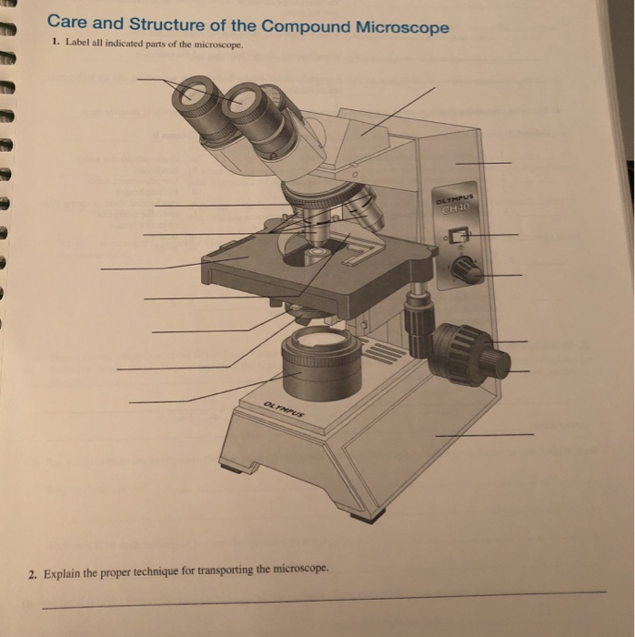
Solved: Care And Structure Of The Compound Microscope 1. L... | Chegg.com
Compound Microscope Parts - Labeled Diagram and their Functions The term "compound" refers to the microscope having more than one lens. Basically, compound microscopes generate magnified images through an aligned pair of the objective lens and the ocular lens. In contrast, "simple microscopes" have only one convex lens and function more like glass magnifiers.

Compound Microscope Diagram | Microscope | Pinterest | Teaching cells, Life science and Cell biology
Diagram of a Compound Microscope - Biology Discussion A bright-field or compound microscope is primarily used to enlarge or magnify the image of the object that is being viewed, which can not otherwise be seen by the naked eye. Magnification may be defined as the degree of enlargement of the image of an object provided by the microscope.
37 Microscope Label And Functions - Understandingluan
PDF Basic Observation Procedures for Compound Microscopes 3. Rotate the 100X objective into position without getting the 40X objective in the oil. 4. While observing from one side of the stage, slowly, raise the stage until you see the meniscus of the oil on the specimen come in contact with the tip of the 100X objective. Now go to the eyepieces and observe as you finish focusing with the fine focus knob.
"Compound Microscope with labels" Stock image and royalty-free vector files on Fotolia.com - Pic ...
› books › NBK26880Looking at the Structure of Cells in the Microscope ... A special sample holder is used to keep this hydrated specimen at -160°C in the vacuum of the microscope, where it can be viewed directly without fixation, staining, or drying. Unlike negative staining, in which what is seen is the envelope of stain exclusion around the particle, hydrated cryoelectron microscopy produces an image from the ...

Sketch the onion peel cell as seen under the microscope. Label the parts such as the cell wall ...
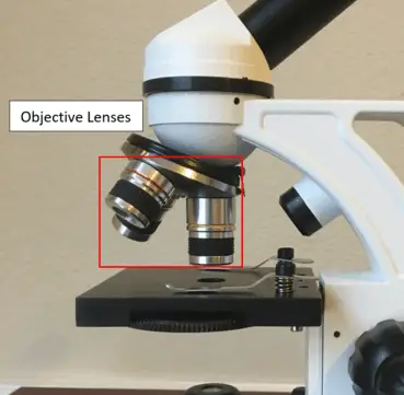
Microscope, Microscope Parts, Labeled Diagram, and Functions Revolving Nosepiece or Turret: Turret is the part of the microscope that holds two or multiple objective lenses and helps to rotate objective lenses and also helps to easily change power. Objective Lenses: Three are 3 or 4 objective lenses on a microscope. The objective lenses almost always consist of 4x, 10x, 40x and 100x powers. The most common eyepiece lens is 10x and when it coupled with ...
template
Compound Microscope Parts Made Easy List and Diagram of Compound Microscope Parts: Head - The head is the uppermost part of the microscope that contains the eyepiece, tube, objective lens, and nosepiece. So all the optical parts of a compound microscope are in the head. Eyepiece - The eyepiece is the lens at the top, and the part you look through when using the microscope.

Help with compound microscope? | Yahoo Answers
What is a Compound Microscope? - New York Microscope Company What is a Compound Microscope? A compound microscope is an instrument that is used to view magnified images of small specimens on a glass slide. It can achieve higher levels of magnification than stereo or other low power microscopes and reduce chromatic aberration. It achieves this through the use of two or more lenses in the objective and the ...
Post a Comment for "42 compound microscope diagram without labels"